NanoPhotometer®: Small Volume Spectrophotometric Applications in Physiology
NanoPhotometer® Application Leaflets
Small Volume Spectrophotometric Applications in PHYSIOLOGY
Myocardial fibrosis and apoptosis in long-term experimental diabetes mellitus
INTRODUCTION:
Diabetes mellitus (DM) is becoming a pandemic. In 2003, 194 million people had diabetes, and this number is predicted to increase by 72%, affecting 366 million in 2030. Of them, at least 10% have type 1 diabetes (DM1). More than 65% of diabetic patients die because of cardiovascular complications. Cardiac damage can be directly induced by DM and enhanced by the coexistence of coronary artery disease and hypertension. The excess of glucose alters metabolic, structural and contractile proteins and activates cellular responses. Although the key processes in DM are known such as as oxidative damage, cardiac cell apoptosis, interstitial fibrosis and an increased reactive oxygen species (ROS) generation, there is much less information on features such as local inflammation. The molecular mechanisms involved in these pathogenic processes are not fully elucidated, and there is not enough information on the long-term injury of the DM1 heart and its relationship with coexistent hypertension. In this sense, the fibrotic, apoptotic and inflammatory events in long- and short-term streptozotocin (STZ)-induced DM1, hypertensive and DM1/ hypertensive myocardium of rats have been studied. The Implen NanoPhotometer®, with its integrated NanoVolume and cuvette capability in addition to a diverse methodical spectrum, was routinely used for important methodical steps in this project. The qualitative and quantitative measure of extracted RNA with the submicroliter cell and the protein quantification in standard cuvettes (BCA assay) could be perfectly performed by this instrument. The fast and constant readings underline the reliable performance of the Implen NanoPhotometer® for nucleic acid and protein analysis.
METHODS:
RNA quantification and QPCR
As described in detail by Area-Carrasco et al. 2009, normotensive and spontaneously hypertensive rats (SHR), DM1/SHR and control (normotensive) male rats were killed after a 6 week (short term DM1) or 22 week (long-term DM1) treatment and the left ventricle isolated. The analysis of relative gene expression was performed by retro-transcription and quantitative PCR (QPCR). Total RNA from left myocardium or cultured cells was extracted and dissolved in TRIzol reagent. Then a white precipitate corresponding to the total tissue/cell RNA was obtained, which could be dissolved in TE buffer. A very small volume of 1 µl of each sample was used for RNA quantification by the Implen NanoPhotometer®, measuring the absorbance at 260 nm. The 260/280 abs. ratios between 1.75 and 2 indicated the purity of the samples. Equal amounts of total RNA were reverse transcribed to obtain the cDNA, which was used for multiplex QPCR (Area-Carrasco et al. 2009).
Protein quantificatifon and Western
Protein extraction and quantification was performed as described in detail by Area-Carrasco et al. 2009. The total protein extract was separated from the supernatant and quantified by the Implen NanoPhotometer®, using the BCA method in standard cuvettes (Abs 562 nm) and BSA as protein standard. For Western Blot assays, equal amounts of protein samples (40 µg) were loaded on an acrylamid gel and transfected to nitrocellulose membranes. The membranes were blocked and incubated with the primary antibodies. Next, membranes were incubated with peroxidase-labelled secondary antibodies and developed with ECL/X-ray films. a-Tubulin and GAPDH were the loading controls (Area-Carrasco et al. 2009).
RESULTS:
Myocardial fibrosis in long-term DM1 and SHR
For the characterization of myocardial fibrosis, induced by long-term STZ-stimulated DM1 and hypertension, morphological changes in the myocardial structure and mRNA expression of fibronectin (FN) as extracellular matrix glycoprotein were studied. Both, DM1 and DM1/SHR rats presented an increased extracellular matrix protein composition vs. control (Figure 1, see red staining).
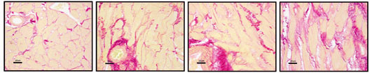
Figure 1: Morphological changes in long-term type 1 diabetes mellitus (DM1) and hypertensive myocardium. Staining with Sirius red in STZ-induced DM1, SHR, DM1/SHR and control hearts.
In DM1 and DM1/SHR myocardium, the fibrosis matrix component (fibronectin) mRNA expression was elevated 1.8-2.3-fold respectively vs. control (Figure 2). Interestingly, SHR rats showed similar fibrosis and FN mRNA expression as DM1/SHR (Figure 1/2).
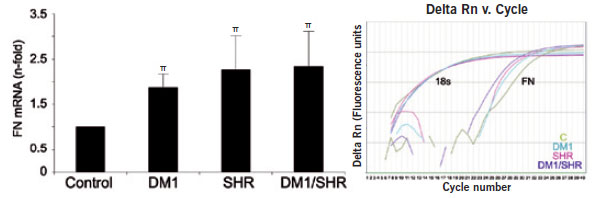
Figure 2: Fibronectin (FN) mRNA expression. A representative QPCR amplification [delta normalized receptor (Rn) vs. cycle] of a rat of each group (control, DM1, SHR, DM1/SHR) for 18 s and FN is also shown. *P < 0.01 or pP< 0.05 vs. control.
CTGF in long-term DM1 and SHR myocardium
The transforming growth factor-ß (TGF-ß) system is a key mediator of fibrosis in the heart and other tissues through the decomposition of extracellular matrix proteins, such as FN and type I collagen from local cells. Connective tissue growth factor (CTGF) is a well-known mediator of the TGF-ß system actions, although its expression has not been described in DM. Here it can be reported that the CTGF mRNA expression was increased in long-standing DM1, SHR and in DM1/SHR myocardium (3.6-, 4.2-, and 3.5-fold, respectively, vs. control mRNA expression, Figure 3).
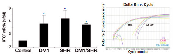
Figure 3: Connective tissue growth factor (CTGF) mRNA expression. A representative QPCR ampli-fication (deta Rn vs. cycle) of a rat of each group (control, DM1, SHR, DM1/SHR) for 18s and CTGF is also shown. pP < 0.05 vs. control.
Long-term DM1, hypertension and cardiac apoptosis
TNF superfamily proteins, such as FasL promote apoptosis through caspase activation. The expression of FasL (Figure 4) and its receptor Fas (data not shown) was increased in DM1 and DM1/SHR myocardium, indicating apoptotic mechanisms in long-term DM1.
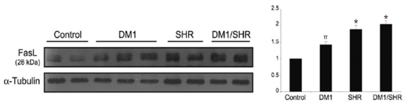
Figure 4: DM1, hypertension and cardiac apoptosis. Myocardial Fas ligand (FasL) levels by Western Blot with a-Tubulin as loading control. Semiquantification of apoptosis (n-folds) for each group is also shown. *P < 0.01 or pP< 0.05 vs. control.
Inflammation in long-term DM1 and SHR myocardium
In the past the myocardial inflammation has been described in SHR and short-term STZ-induced DM1 by an increased expression of pro- inflammatory cytokines like IL-1ß and TNF-a. However, in long-term DM1, MCP-1, VCAM-1 and TNF-a (Figure 5 and 6A) as well as IL-1ß and IL-6 (data not shown) were significantly elevated.
It could be confirmed that myocardial pro-inflammatory factors are increased in hypertension (Figure 5/6) and short-term DM1 (data not shown), in correlation with the presence of inflammatory cells (data not shown). Interestingly, in long-term DM1 the local expression of anti- inflammatory and antioxidant molecules (Figure 6B) may underline the absence of the inflammatory recruitment (not shown).
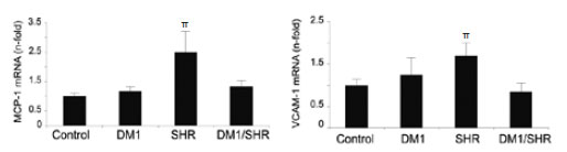
Figure 5: MCP-1 and VCAM-1 mRNA expression of rats of each group (control, DM1, SHR, DM1/SHR). *P < 0.01 or pP< 0.05 vs. control.
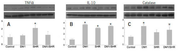
Figure 6: DM1, hypertension and inflammation. Myocardial cytokine level of proinflammatory (A) and anti-inflammatory/antioxidant IL-10 (B) and Catalase (C) by Western Blot with GAPDH as loading control. Semiquantification (n-folds) for each group (control, DM1, SHR, DM1/SHR) is also shown. *P < 0.01 or pP< 0.05 vs. control.
In long-term DM1, many features of short-term myocardial injury are maintained. However, the inflammatory process appears to be blunted, possibly due to the local expression of anti-inflammatory and antioxidant molecules. Moreover, at this stage, the effect of both, combined DM1 and hypertension are, for the most part, not additive.
A. Ortiz, J. Egido, J. Tunon and O. Lorenzo. Myocardial fibrosis and apoptosis, but not inflammation, are present in long-term experimental diabetes. Am. J. Physiol. Heart Circ. 297: H2109-H2119, 2009.
SUMMARY:
Diabetes mellitus (DM) is the most popular metabolic disease of the human species. In 2010, 6.4% of the human population had diabetes and based on international evaluations, the numbers will dramatically rise during the next decades. Besides the defective function of the insulin system, there are many patholo-gical mechanisms connected with DM. Cardiac damage is the most common results of malfunctions in the insulin balance, and can be enhanced by the coexistence of coronary artery disease and hypertension. Hypertrophy, fibrosis, apoptosis and inflammation have been described in early stages of experimental DM1 myocardial injury. However, there is still not enough information on the long-term injury of the DM1 heart and its relationship with coexistent hypertension. Therefore, investigations of fibrotic, apoptotic and inflammatory events in long- and short-term DM1, hypertensive, and DM1/hypertensive myocardium are of great interest.
The present studies show that the combination of DM1 and hypertension results in non-significant additive effects. Moreover, the coexistence of DM1 blunted the inflammatory response to hypertension. Interestingly, anti-inflammatory IL-10 and anti- oxidants were induced in long-term DM1 and DM1/SHR hearts. And although fibrosis and apoptosis are features of long-term myocardial damage in experimental DM1, associated hypertension does not induce additional changes. On the other hand, myocardial inflammation is present in hypertension and short-term DM1, but is not a key feature in long-term DM1.
For the investigations of different parameters involved in long-term DM1, the Implen NanoPhotometer® played an important role for many methodical steps. Concentrations of RNA and proteins could be easily and accurately determined using the NanoVolume and cuvette options. The multifunctional, maintenance-free NanoPhotometer® simplifies the effort
We would like to thank Dr. Óscar Lorenzo, Vascular Pathology and Cardiology Department, Autónoma University of Madrid, Spain, for the friendly offer of measuring data, and for supporting us during the compilation of this Physiology application leaflet!